Friday, November 23, 2018


Klenow Fragment
It is a large fragment of DNA polymerase I enzyme. It results from the proteolytic cleavage, of DNA polymerase I, caused by the protease subtilisin. Of the 3 main purposes of DNA polymerase I, the Klenow fragment possesses two of them, which are:
·         5’ 3’ polymerase activity.
·         3’ 5’ exonuclease activity.
So basically, the Klenow fragment is a DNA polymerase I enzyme, without the 5’ 3’ exonuclease activity. But having those two properties makes the Klenow fragment have applications such as:
®     The preparation of radioactive DNA probes.
®     DNA sequencing through the Sanger method.
®     Synthesis of second strand of cDNA.


Reference:
Sambrook, J., Fritsch, E.F. and Maniatis, T. (1989). Molecular Cloning: A Laboratory Manual. (2nd ed.), 5.40-5.43. Cold Spring Harbor: Cold Spring Harbor Laboratory Press.

T4 Polynucleotide Kinase


Ø  Definition:
T4 Polynucleotide Kinase catalyzes the transfer of a phosphate from ATP to the 5´-terminus of polynucleotides or to mononucleotides bearing a 5´-hydroxyl group. The enzyme may be used to phosphorylate RNA, DNA and synthetic oligonucleotides prior to subsequent manipulations such as ligation and cloning.
ATP + 5'-dephospho-DNA {\displaystyle \rightleftharpoons } ADP + 5'-phospho-DNA        

Ø  Applications:
The T4 polynucleotide kinase is used for labeling the 5'-termini of nucleic acids, and the labeled products can be used int he following ways: - markers for gel-electrophoresis - primers for DNA sequencing - primers for PCR - probes for hybridization - substrates for DNA and RNA ligases - probes for transcrip mapping 5'-phosphorilation of oligonucleotide linkers and DNA or RNA is required before ligation can be performed. using the 32P-postlabeling assay, DNA modification can be detected.

Ø  Source:
 T4 PNK is purified from E. coli cells expressing a recombinant clone

Ø  Common reaction buffer:
The buffer contains 70 mM Tris-HCl, 10 mM MgCl2, 5 mM DTT and pH 7.6 at 25°C

Ø  Structure:


Structure of the 5′kinase domain. Two enzymebound sulfates coordinated at the active site tunnel are shown as a stick models. Coordination of one sulfate within the oxyanion hole formed by the Ploop is depicted by dashed lines.

Ø  References
https://worldwide.promega.com/products/cloning-and-dna-markers/molecular-biology-enzymes-and-reagents/t4-polynucleotide-kinase/?catNum=M4101

By:
·       Adrian Barnutiu A86084
·       Carlos Rodrigues A85152
·       Catarina Deseyve A85111
·       Gabriel Abreu A84397
·       Matilde Rocha A85740

Enzymes that use DNA as substrate- Nuclease S1


S1 nuclease is a restriction enzyme, specifically an endonuclease (enzyme capable of cleaving the DNA strand at specific sites) derived from the organism Aspergillus oryzae (a fungus).
S1 nuclease is normally present in fungi and plants and is stable at 65°C.
The chemical reaction catalyzed by S1 is an endonucleolytic cleavage that results in 5'-phosphomononucleotides and 5'-phospho-oligonucleotides, requiring Zn2+ as the cofactor.
The main function of this enzyme is to remove protrusions in ssDNA (single stranded DNA) and to divide the DNA or RNA (targets preferencially DNA) into oligonucleotides, in order to facilitate the work in molecular biology.
The common reaction buffer is a solution of 10 mM sodium acetate (pH 4.6), 150 mM NaCl, 0.05 mM ZnSO4 and 50% glycerol.
Double-stranded nucleic acids can resist degradation by S1 nuclease except when concentration is extremely high.


Cristia Pires- A94335
Filipa Sampaio-A81577
Marta Machado-A81723
  Marta Mendanha - A82382
 Mónica Fernandes - A84741

Enzymes that use DNA as a substrate - Bam HI

Enzymes that use DNA as a substrate

Bam HI is a restriction endonuclease and that means it cleaves the DNA in a specific sequence of nucleotides. Was isolated from a microorganism, more precisely from Bacillus amyloliquefaciens. This enzyme goes through all DNA molecule and then, when recognizes the sequence GGATCC and cleaves the molecule between the two guanine nucleotides.

Bam HI is more effective at 37ºC and isn’t sensitive to heat inactivation. It is storage at -20ºC in this conditions: (10 mM Tris-HCl; 50 mM KCl; 1 mM DTT; 0.1 mM EDTA; 200 μg/ml BSA; 50% Glycerol; pH 7.4 @ 25°C). Also, Bam HI can cut substrate DNA in 5-15 minutes and can be digested overnight without destroying the DNA.

The principal applications of Bam HI are molecular cloning, restriction site mapping, Genotyping, Restriction fragment length polymorphism (RFLP) and single nucleotide polymorphism (SNP).

The buffer used to incubate the Bam HI is different from company to company, but one of them is: (100 mM NaCl;50 mM Tris-HCl;10 mM MgCl2;100 μg/ml BSA ;(pH 7.9 @ 25°C))

References:
New England Biolabs.inc:
            ThermoFisher scientific:
            Protein sequence analysis and classification:


Grupo2.3: Sofia Salgado, Simão Ferreira, Tânia Castro, Tiago Ribeiro, Eduardo Faia

Applied Biology 2nd year, University of Minho


AluI


Definition (reaction and biologic origin):
AluI is a restriction enzyme (protein that cuts DNA into fragments according to specific nucleotide sequences). It recognizes the sequence AGCT and cuts between the G and C nucleotides.
This enzyme is extracted from E. coli cells whose DNA was modified to carry the gene AluI from Arthrobacter luteus, which is a bacteria commonly found in the soil.

Uses:
   AluI is used in: molecular cloning, restriction site mapping (method used to map an unknown segment of DNA by breaking it into pieces and then identifying the locations of the cuts), genotyping (determines differences in genomes by comparing a DNA sequence to that of another sample or a reference sequence, and it can identifie small variations within populations), restriction fragment length polymorphism (RFLP) and single nucleotide polymorphism (SNP).

Buffer:
The buffer used in reactions with AluI is Tango buffer which is premixed with BSA (Bovin Serum Albumin, a protein derivated from cows that gives stability to the enzymes and binds with contaminants that may be presente in DNA preparations) and ensures the optimum reaction conditions for restriction enzymes.

References:

Unknown author. Date unknown. AluI. Retrieved on November 14, 2018. Available at: https://international.neb.com/products/r0137-alui#Product%20Information
Unknown author. Date unknown. AluI restriction enzyme. Retrieved on November 14, 2018. Available at: https://www.takarabio.com/products/cloning/restriction-enzymes/alui
Unknown author. Date unknown. AluI (10 U/µL). Retrieved on November 15, 2018. Available at: https://www.thermofisher.com/order/catalog/product/ER0011

Unknown author. 2014. Restriction mapping. Retrieved on November 15, 2018. Available at: https://www.nature.com/scitable/definition/restriction-mapping-283

Thursday, November 22, 2018


Restriction enzymes: Sau3AI

Restriction enzymes, also known as endonucleases, are a type of proteins that cleave the phosphodiester bonds of DNA chains at or near a specific recognition site (the sequence of nucleotides is located by the enzyme itself). These enzymes can be isolated from the bacterial cells that produce them and then used to manipulate fragments of DNA (that may contain genes). For this reason, restriction enzymes are indispensable tools for recombinant DNA technology and genetic engeneering. Their applications include molecular cloning, genotyping, RFLP, SNP, etc.
Sau3AI is an example of a type II restriction enzyme, cutting DNA close or within the nucleotide sequence. In this case, the nucleotide sequence recognized by the enzyme is 5’GATC3’ , cleaving the DNA chain before the G (guanine) nucleotide. Its optimal reaction temperature is 37ºC and it should be stored at -20ºC to prevent its activity.


                                 Figure 1- Crystal structure of the C-terminal of Sau3AI fragment.


References:


Enzymes that use DNA as a substract - DNA Polymerase I

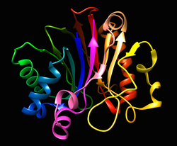
Figure 1 - Functional domains in the Klenow Fragment (left)
and DNA Polymerase I (right).

DNA polymerase I is an enzyme that participates in the DNA replication of prokaryotes. It is the predominant and most abundant polymerizing enzyme found in E.coli. It was the first DNA polymerase to be identified and discovered by Arthur Kornberg, an American biochemist in 1956. He was able to characterize the enzyme initially in E.coli. In E.coli the enzyme is a single polypeptide chain comprised of 928 aminoacids and it has a molecular weight of 109kDa. Nevertheless, the enzyme does not occur only in E.coli but also in many prokaryotes. In prokaryotes it is mostly encoded by polA gene.


Uses:

It has three distinct functions: 3′ to 5′ exonuclease or “proofreading” function because it can remove a nucleotide from the 3’ terminus which was incorrectly inserted in the DNA strand, 5′ to 3′ exonuclease and 5′ to 3′ polymerase.
The second function is the 5’ to 3’ exonuclease. This enzyme has the ability to remove nucleotides from the 5’ terminus. Furthermore, the nucleotides removed can be either of the deoxyribonucleotides or ribonucleotides. The main goal of the 5′ to 3′ exonuclease activity is to remove ribonucleotide primers that are used in DNA replication.
The 5’ to 3’ polymerase is the main activity of the enzyme and it means that it can add nucleotides to the 3’-OH terminus of the previous nucleotide in a DNA template. In order for polymerase to start adding nucleotides it needs a primer (consisted of approximately 10 ribonucleotides) to begin DNA synthesis. DNA pol I adds approximately 15-20 nucleotides per second.


Common reaction buffer:

A common reaction buffer is Tris-HCl (pH 7.2).


References:

ScienceDirect, DNA Polymerases,, viewed in November 13, 2018, https://www.sciencedirect.com/topics/neuroscience/dna-polymerase-i

Worthington Biochemical Corporation, DNA Polymerase I, viewed in November 13, 2018, http://www.worthington-biochem.com/DNAECI/default.html

Biology Online, DNA Polymerase I, viewed in November 13, 2018, https://www.biology-online.org/dictionary/DNA_polymerase_I

Technical information of kit M205A, DNA polymerase I, Promega Corporation.


Evangelia Nikou (e8749) | Hugo Rodrigues (a85946) | Mélanie Pereira (a83980) 
Pedro Gonçalves (a84784) | Ricardo Fernandes (a86254)

University of Minho | School of Sciences | Department of Biology 

Degree in Applied Biology | Genes and Genomes

Monday, November 19, 2018

Enzymes that use DNA as a substrate - Deoxyribonuclease I (DNASE I)


Deoxyribonuclease I (DNAse I) is a versatile enzyme (endonuclease) that catalyzes the hydrolytic cleavage of the adjacent phosphodiester bonds of pyrimidines nucleotides (deoxycytidine and deoxythymidine), causing these bonds to degrade, releasing di/tri/oligo phosphorylated nucleotides as products. It is coded by the human DNASE1 gene.


It is a powerful research tool in the field of molecular biology for DNA manipulation. It can be used:
  • To create a library of fragments of  DNA sequences for in vitro use of recombination reactions. 
  • In the degradation of contaminated DNA after RNA isolation;
  • In the identification of the protein binding sequence in DNA (DNAse I footprinting).
    Resultado de imagem para DNAse1
    Figure 1 DNAse I  diagram.


The most common buffer components for the use of DNAse I are 10mM Tris-HCl; 2.5 mM MgCl2; 0.5mM CaCl2; pH 7.6 at 25ºC, and we must store this solution at -20ºC.


References:

R. A. Bowen; Laura Austgen; Melissa Rouge (2000). "Restriction Endonucleases and DNA Modifying Enzymes". Nucleases: DNase and RNase. Biotechnology and Genetic Engineering.


National Center for Biotechnology Information, U.S. National Library of Medicine (2018). DNASE1 deoxyribonuclease 1 [Homo sapiens (human)]. Retrieved at november 13 2018, from: https://www.ncbi.nlm.nih.gov/gene/1773


Thermo Fisher Scientific. (2016). DNAse I Demystified. Retrieved at november 13 2018, from: https://www.thermofisher.com/pt/en/home/references/ambion-tech-support/nuclease-enzymes/general-articles/dnase-i-demystified.html


New England Biolabs. (2018). DNAse Reaction Buffer. Retrieved at november 13 2018, from:  https://international.neb.com/products/b0303-dnase-i-reaction-buffer


Jazzlw. (2016). DNAse I. Retrieved at november 13 2018, from: https://commons.wikimedia.org/w/index.php?curid=47824954


Genes e Genomas | Biologia Aplicada | Universidade do Minho
A86073 Ana Beatriz Maia | A85219 Ana Rita Campos
A84133 Jorge Gonçalves | A83512 Vera Oliveira

Enzymes that use DNA as substrate: Alkaline phosphatase _ group 3.2

The Alkaline phosphatase (ALP) is an enzyme that catalyzes dephosphorylation of compounds at basic pH values (catalyzes the hydrolysis of monoesters in phosphoric acid) which can additionally catalyze the transphosphorylation reaction with large concentrations of phosphate acceptors.
The enzyme is found in prokaryotes and eukaryotes with the same general function but in different structural forms suitable to the environment they function in. In E. coli, alkaline phosphatase is found in the periplasmic space while in humans is found in bile ducts, bone, intestine, placenta, and tumors.


Typical uses in the lab for alkaline phosphatases include: removing the phosphates groups on the 5' end to prevent the DNA from ligating (the 5' end attaching to the 3' end), thereby keeping DNA molecules linear until the next step of the process for which they are being prepared; also, removal of the phosphate groups allows radiolabeling (replacement by radioactive phosphate groups) in order to measure the presence of the labeled DNA through further steps in the process or experiment.

                                           Figure 1: Alkaline phosphatase reaction.

The common reaction buffer used is the Alkaline phosphatase 10X Reaction Buffer (M183A).


Bibliography

·         Telega, Grzegorz W.,Learn more about Alkaline phosphatase,2018. Disponível em: https://www.sciencedirect.com/topics/neuroscience/alkaline-phosphatase. Acesso em 13 de novembro de 2018.
·         Sharma, Ujjawal; Pal, Deeksha; Prasad, Rajendra; Alkaline Phosphatase: An Overview, 2014. Disponível em: https://www.ncbi.nlm.nih.gov/pmc/articles/PMC4062654/. Acesso em 13 de novembro de 2018.
·         Phosphatase, Alkaline. Disponível em: http://www.worthington-biochem.com/bap/default.html. Acesso em 13 de novembro de 2018.
·         Disponível em: https://www.promega.com/-/media/files/resources/protocols/product-information-sheets/g/alkaline-phosphatase-calf-intestinal-protocol.pdf. Acesso em 13 de novembro de 2018.


Ana Rita Pacheco (a83934) | Angelina Eiras (a80415) | Margarida Lima (a84229) | Maria Freitas (a84345) | Maria Rui Ferreira (a85952)  

University of Minho | School of Sciences | Department of Biology  
Degree in Aplicated Biology | Genes and Genomes

Pancreatic RNase A


   The Pancreatic RNase A is a pyrimidine-specific endonuclease found in the pancreas of certain mammals and of some reptiles. Since this is an endonuclease that means the function of this enzyme is to degrade phosphodiester bonds.

    This enzyme is usually extracted from Bovine Pancreas, or scientists extract the enzyme using a technic called recombinant DNA. In this case, they take the gene from the pancreas of a mammal and them inserte that gene in the plasmid of an mutante strain of E. Coli.

Functions:
-  Removal of RNA for RNA free DNA purification reactions such as plasmid DNA purification and genomic DNA purification;
- RNA removal from recombinant protein preparations;
-Ribonuclease protection assays;
-Mapping single-base mutations in DNA/RNA;
- RNA sequence analysis and protection assays.

The reaction buffer is Tri-HCl, pH 7.5.

                                                  Figure 1: Structure of RNase A

References:

      
·    Work done by Group 3.4: Henrique Sousa - A84794; Marco Malheiro - A85487; Ricardo Alves - A83636; Vítor Alves - A86253
        
     Applied Biology 2nd year 2018/2019 - University of Minho – November 19th, 2018

Friday, November 2, 2018


Analysis of Saccharomyces cerevisiae’s chromosome X

The final sequence of Saccaromyces cerevisiae‘s genome was assembled from roughly 300,000 independent sequence reads, with error rates from 0.5 to 1%, resulting in an estimated error rate of the final sequence of less than 3 errors in 10,000 bases (0.03%). The complete sequence was made available to the public on April 24th, 1996.
Only 43.3 % of the yeast genes are currently classified as ‘functionally characterized’, having experimentally well-investigated properties, being members of well-defined protein families, or displaying strong homology to proteins with known biochemical functions.
The size of the genome is approximately 12071326 bp.

The chromosome X has a length of 7745442 bp, having each arm a length of 436307 bp and 309017 bp, and the total number of functional genes (ORF – Open Reading Frame) is 379. (The complete nucleotide sequence of ARS 1025 is illustrated on Figure 1).
It contains 25 Autonomously Replicating Sequences (ARS) and five of them are listed, along with their functions, on Table 1.


Table 1- Genes involved in replication

References:
·         F.Galibert, et al., 1996, Complete nucleotide sequence of Saccharomyces cerevisiae chromosome X, EMBO J 15(9), page 1-2;
·         SGD: Saccaromyces cerevisiae Genome Snapshot – www.yeastgenome.org/genomesnapshot - October 29th, 15h30min.

Work carried by Group 3.3: João Cruz, José Freitas, Margarida Barros and Mariana Pereira
Biochemistry 2nd year 2018/2019 - University of Minho – October 29th, 2018

Thursday, November 1, 2018

Chromosome 2 (Saccharomyces cerevisiae)


Chromosome 2 (Saccharomyces cerevisiae)


    The DNA sequence of the yeast Saccharomyces cerevisiae chromosome II has 807 188 bp, whole the left arm has 238207 bp and the right arm has 575579 bp.  At present, it is the largest eukaryotic chromosome entirely sequenced.  This chromosome contains a total of 410 functional genes (covering 72% of the sequence). Searches revealed that 124 ORFs (30%) correspond to genes of known function, 51 ORFs (12.5%) appear to be homologues of genes whose functions are known, 52 others (12.5%) have the functions of which are not well defined and another 33 of the novel putative genes (8%).
This chromosome presents 21 ARS, such as: ARS201; ARS230; ARS202; ARS203; ARS206; of which the ARS203 (figure 1) is involved in replication.
In summary, Chromosome II has various genes that are responsible for very different functions, as you can see in the next link: https://wiki.yeastgenome.org/images/c/c4/Chr2.pdf

Figure 1- Sequence of the ARS203.


Grupo 1.2
Beatriz Ferreira
Diana Alves
Filipe Guimarães
Francisca Gonçalves
Samuel Rocha

2º ano Licenciatura em Bioquímica | Universidade do Minho